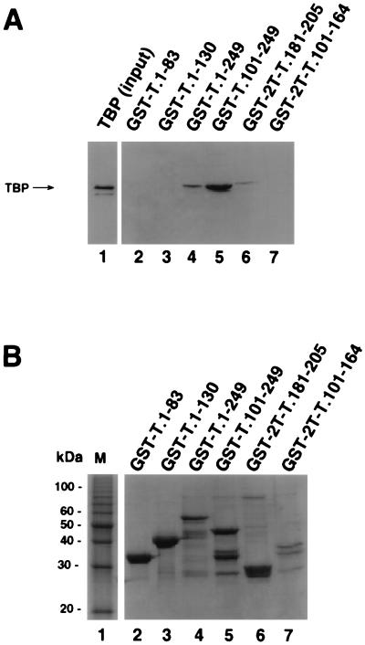
FIG. 2.
A 24-amino-acid region of T antigen is sufficient for specific interaction with TBP. (A) Purified full-length TBP was incubated with beads containing the indicated GST-T antigen fusion proteins (lanes 2 to 7) as described for Fig. 1. After the beads were washed, the bound proteins were analyzed by SDS-PAGE (12.5% gel), and TBP was detected by immunoblotting using the monoclonal anti-TBP antibody 4C8. Lane 1 contained 0.1 μg of the input TBP. (B) The GST-fusion proteins used for A (10 μl) were analyzed by SDS-PAGE (12.5% polyacrylamide gel) and staining with Coomassie brilliant blue. M, 10-kDa marker protein ladder.

