Figure 3.
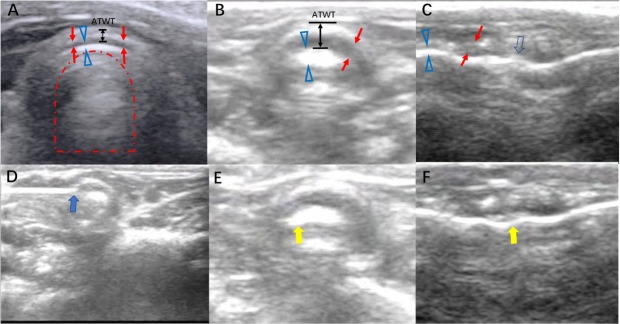
Sonogram of rabbit trachea and the procedure of real-time guidance of transcervical ultrasound for submucosal paclitaxel administration for tracheal stenosis (A) Sonogram of the normal trachea by transcervical ultrasound in the transverse plane. Red arrow, as a hypoechoic shadow, indicates the tracheal ring; hollow blue triangle, as a hyperechoic shadow, indicates the tissue–air border, named the A-M interface; red dotted line arch indicates the air artifact in the trachea; black double arrow line indicates the ATWT. (B) Sonogram of tracheal stenosis by transcervical ultrasound in the transverse plane. (C) Sonogram of tracheal stenosis by midsagittal plane scan. The hollow blue arrow indicates an unsmooth A-M interface, corresponding to the irregularity of the inner side of the trachea. (D) Injection of paclitaxel by a 25G needle into the submucosal site of the trachea (blue solid arrow). (E, F) After the submucosal injection of paclitaxel, transcervical ultrasound revealed a change in the A-M interface (yellow solid arrow), indicating the administration of paclitaxel to the submucosal layer of trachea. A-M: air-mucosal; ATWT: anterior tracheal wall thickness.

