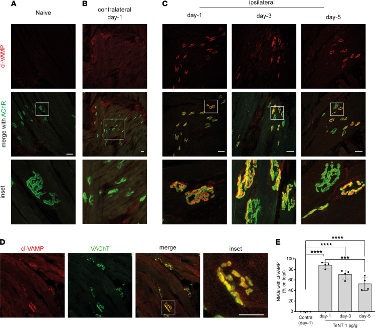
Figure 2. TeNT cleaves its target VAMP at motor axon terminals of the WP in mice.
Confocal images of WP musculature from (A) naive and TeNT-treated mice (B) contralateral or (C) ipsilateral to injection at indicated times after injection; the red signal indicates the cleavage of VAMP at the NMJ identified through the labeling of nicotinic acetylcholine receptors (AChR, shown in green) with fluorescent α-bungarotoxin; insets show a 5× original magnification (A and C) and 3× original magnification in B. (D) Confocal images showing colocalization between cl-VAMP (red) and the vesicular transporter of acetylcholine (VAChT, green), a protein marker of synaptic vesicles, as expected from TeNT cleavage of VAMP on synaptic vesicles at the motor axon terminal; scale bar, 50 μm. (E) Quantification reporting the percentage of NMJs positive for the signal of cl-VAMP in the ipsilateral WP at indicated time points after TeNT injection compared with the contralateral at day 1. Data are expressed as means ± SD; ***P < 0.001; ****P < 0.0001 assessed by 1-way ANOVA with Bonferroni’s test. Black circles indicate the number of animals used in the experiment.

