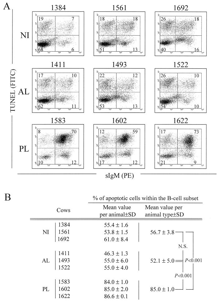
FIG. 3.
Detection of apoptosis in B lymphocytes from BLV-infected PL (no. 1583, 1602, and 1622), BLV-infected AL (no. 1411, 1493, and 1522) and NI (no. 1384, 1561, and 1692) cows. PBMCs were cultured for 24 h and labeled with the PIg45A2 MAb (directed against bovine IgM) and a phycoerythrin (PE)-conjugated secondary antibody. The cells were then fixed and processed by the TUNEL procedure to detect apoptosis, and samples were then analyzed by dual-immunofluorescence flow cytometric analysis. (A) Results from a representative experiment are presented as dot plots. On the basis of control staining, each distribution was divided into four quadrants. The numbers indicate the percentage of PBMCs in each quadrant. (B) Percentages of apoptotic cells within the B-lymphocyte population. The mean values and standard deviations (SD) were calculated with three animals from two independent experiments. The significance of the differences in mean values between groups of animals was established by a Student t test. N.S., not statistically significant.

