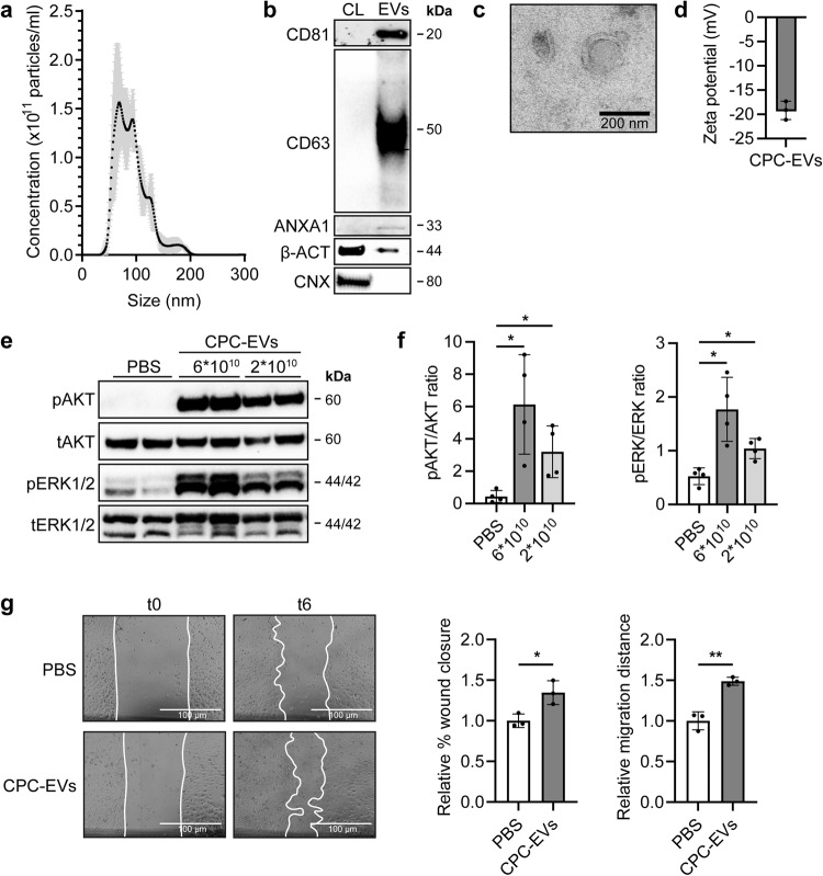
Fig. 1. CPC-EVs activate intracellular signalling in HMEC-1 and induce HMEC-1 migration.
a Representative NTA plot showing the size distribution and particle concentration of EVs isolated from the conditioned medium of CPCs (CPC-EVs) using size-exclusion chromatography. b Western blot analysis showing the presence of CD81, CD63, Annexin A1 (ANXA1), β-actin (β-ACT), and absence of Calnexin (CNX) in CPC-EVs. β-ACT and CNX were present in CPC lysate (CL). c Representative TEM image of CPC-EVs. d Surface charge (zeta potential) of CPC-EVs as measured by laser Doppler electrophoresis. e, f Representative western blot analysis of phosphorylated AKT (pAKT), total AKT (tAKT), phosphorylated ERK1/2 (pERK1/2) and total ERK1/2 (tERK1/2) in HMEC-1 treated with two different CPC-EV particle doses (dosing based on ref. 12). f Quantification of pAKT, tAKT, pERK1/2 and tERK1/2 expression levels using densitometry expressed as pAKT/AKT and pERK/ERK ratios (n = 4). g Wound healing assay showing effects of CPC-EVs on HMEC-1 migration 6 h (t6) after EV addition (t0), analysed both as relative % wound closure and relative absolute migration distance compared to PBS (n = 3). Data are presented as mean ± SD. *p < 0.033; **p < 0.0021.

