. 2023 Jul 25;12(1):A0128. doi: 10.5702/massspectrometry.A0128

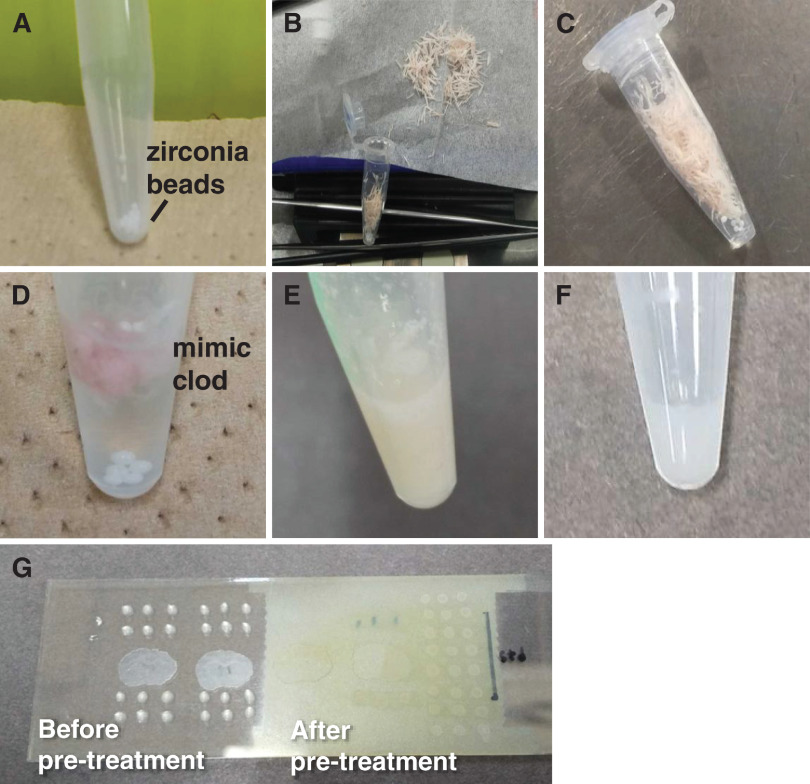
Fig. 1. Sample preparation protocol. (A) Weigh zirconia beads in a 1.5-mL tube. (B) Collect the trimmed pieces. (C) Weigh the collected trimmed pieces. (D) Add 50 µL of ultrapure water to 10 mg of trimmed pieces. (E) Mimic tissue after mixing with a vortex mixer. (F) Mixture of mimic tissue and standard solution. (G) 0.5 µL spot on ITO glass (left), after derivatization reagent and matrix sprayed with an airbrush (right). ITO, indium–tin–oxide.

Fig. 1. Sample preparation protocol. (A) Weigh zirconia beads in a 1.5-mL tube. (B) Collect the trimmed pieces. (C) Weigh the collected trimmed pieces. (D) Add 50 µL of ultrapure water to 10 mg of trimmed pieces. (E) Mimic tissue after mixing with a vortex mixer. (F) Mixture of mimic tissue and standard solution. (G) 0.5 µL spot on ITO glass (left), after derivatization reagent and matrix sprayed with an airbrush (right). ITO, indium–tin–oxide.