FIGURE 1.

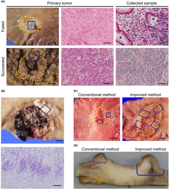
Possible reasons for unsuccessful gastric cancer stem cell (GC‐SC) spheroid culture. (A) Macroscopic luminal views of the resected specimens (left) and H&E‐stained sections of the primary tumors (center) and collected tissue samples (right) in a failed (top) and a succeeded (HG6T, bottom) case. Yellow dotted lines outline the tumor area. Blue boxes show the regions of sample collection. Note that a collected sample of the failed case contains non‐neoplastic glandular epithelial cells (asterisks). Scale bar, 10 mm (left) and 50 μm (center and right). (B) A macroscopic view of a necrotic GC case (top) and a periodic acid–Schiff‐stained section (bottom) of the collected tumor region (top, red box), showing accumulation of fungal hyphae on the surface. The blue box shows another resected region with successful spheroid culture (HG5T). Scale bar, 10 mm (top) and 50 μm (bottom). (C) Macroscopic views of representative GC cases indicating tumor regions for sample collection (blue boxes) before (conventional method, left) and after improving the method (improved method, right). Yellow dotted lines outline the tumor area. Note that wider regions across the tumor boundary were dissected for the improved method. Scale bar, 10 mm. (D) A cross‐sectional view of a representative GC case indicating the depth of tumor dissection for sample collection. Cutting along a dotted line can result in missing cancer cells in the tissue sample (conventional method). The cancer tissue should be cut deeply along a solid line to obtain enough cancer stem cells (improved method). Scale bar, 5 mm.
