FIGURE 4.

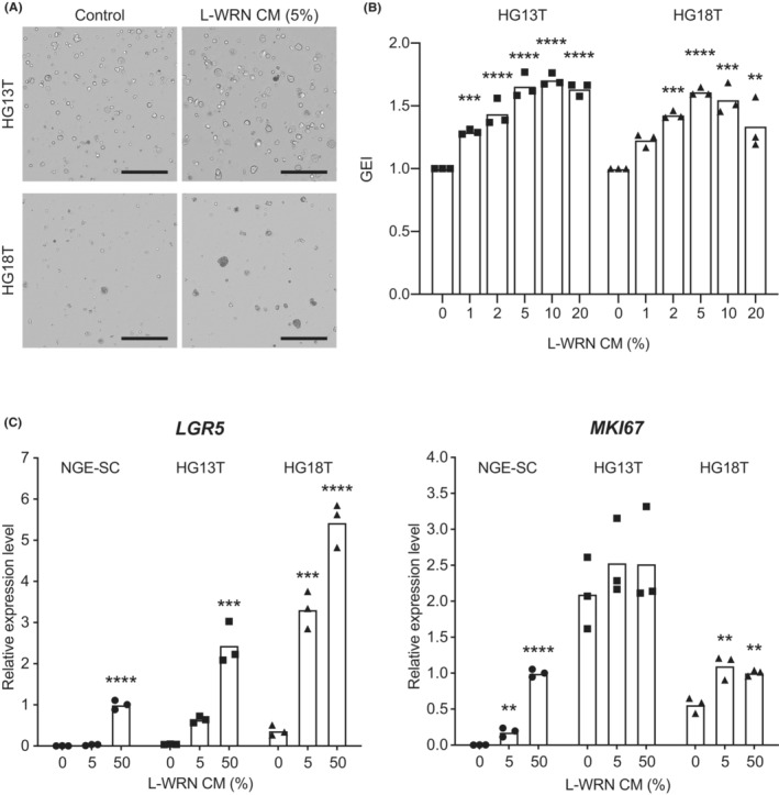
Effects of L‐WRN conditioned medium (CM) on gastric cancer stem cell (GC‐SC) spheroid growth. (A) Representative cell scanning images of HG13T (top) and HG18T (bottom) spheroids cultured with (right) and without (control, left) 5% L‐WRN CM for 6 days. Scale bar, 1 mm. (B) Growth monitoring of HG13T (left) and HG18T (right) spheroids with optical cell imaging. The GEI were calculated based on the growth rate of untreated spheroids (0%). The GEI in three independent experiments are plotted with the means. (C) Expression levels of LGR5 (left) and MKI67 (right) mRNAs determined by quantitative RT‐PCR analysis. Normal gastric epithelial stem cell (NGE‐SC) and GC‐SC (HG13T and HG18T) spheroids were cultured in the cancer media containing 0%, 5%, or 50% L‐WRN CM for 3 days. Relative expression levels in three independent experiments are plotted with the means. **p < 0.01; ***p < 0.001; ****p < 0.0001, statistical significance of the data difference between untreated (0%) and treated groups (one‐way ANOVA followed by Tukey's post‐test).
