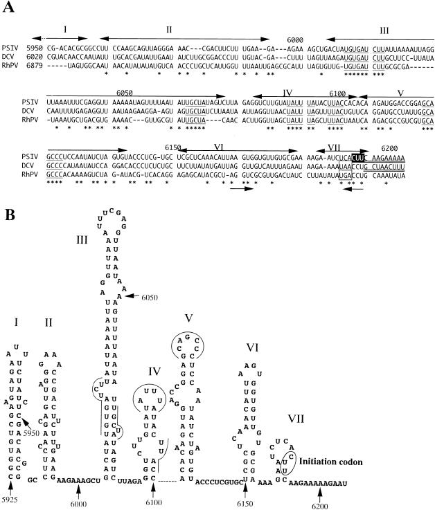
FIG. 7.
(A) Multiple alignment of nucleotide sequences upstream of PSIV (28), DCV (11), and RhPV (19) (accession no. AB006531, AF014388, and AF022937, respectively) capsid-coding regions. The numbers on the left indicate the starting nucleotide positions of the aligned sequences, and the numbers above the sequences represent the nucleotide positions in the PSIV sequence. The initiation codon for the PSIV capsid protein translation is shown in reverse type. In the PSIV and DCV sequences, the capsid-coding regions are doubly underlined. The DCV sequence has a stop codon (boxed) 2 codons upstream of the 5′ terminus of the capsid-coding region, and the RhPV sequence also has a stop codon in the same position. Asterisks indicate nucleotides conserved in all three viruses, and the conserved short RNA segments are underlined. Two arrows below the sequences show an inverted repeat. The double-headed arrows above the sequences represent stem-loop segments in the secondary structure predicted for the PSIV RNA, and the roman numerals correspond to those shown in panel. (B) Computer-predicted secondary structure of the PSIV RNA sequence containing the IRES for the capsid protein translation. The numbers indicate nucleotide positions. The initiation codon is circled. The lines and curves indicate the conserved short RNA segments. Stem-loop structures are numbered from I to VII.

