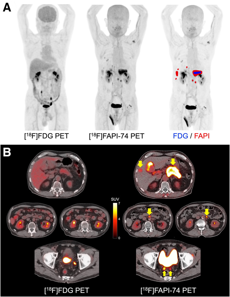
FIGURE 3.
A 68-y-old man with multiple LN and peritoneal metastases of pancreatic cancer. (A) Comparison of maximum-intensity projection images: image on right shows fusion with positive lesions on [18F]FAPI-74 PET (red area) and [18F]FDG PET (blue area). (B) PET/CT images on [18F]FAPI-74 PET and [18F]FDG PET (arrows indicate metastatic lesions). [18F]FAPI-74 PET detected more metastatic lesions than [18F]FDG PET (SUVmax of primary lesion is 9.4 and 3.2, respectively). Brain uptake is decreased on [18F]FDG PET because of high blood glucose level (277 mg/dL).

