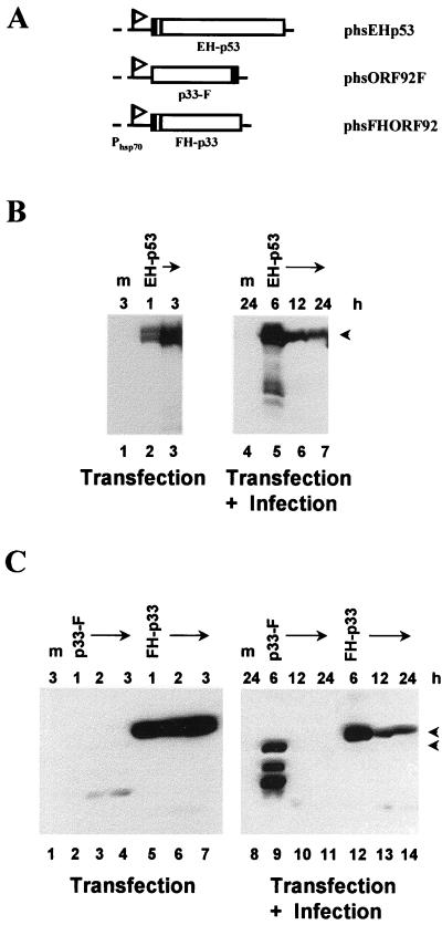
FIG. 2.
Construction and expression of epitope-tagged versions of human wild-type p53 and baculovirus p33. (A) Schematic representation of the plasmids encoding epitope-tagged p53 and p33. The p53 and p33 genes were placed under the transcriptional control of the D. melanogaster Phsp70, indicated by a flag. The Epi and Flag tags are denoted by the filled regions; a sequence encoding six histidines (checkered region) was included in phsEHp53 and phsFHORF92. The identity of each chimeric protein or plasmid is shown below or on the right. (B and C) Immunoblot analysis of the expression of Epi-tagged p53 (B) and Flag-tagged p33 (C). SF-21 cells were mock transfected (m) or transfected with a plasmid expressing the protein indicated above each lane. Cells were harvested after induction by heat shock as indicated above each lane in the left-hand panels. Equal amounts of cell lysates were analyzed by SDS-polyacrylamide gel electrophoresis followed by Western blotting with the mouse anti-Epi monoclonal antibody (B) or with the mouse anti-Flag monoclonal antibody M2 (C). Positions of full-length proteins are indicated by arrowheads on the right. To analyze the expression of tagged p53 and p33 during infection (right-hand panels), transfected SF-21 cells were infected 18 h after transfection and then heat shocked at 6, 12, or 24 h postinfection (as indicated above each lane). Cells were harvested 3 h after heat shock, and equal amounts of cell lysates were analyzed as described above.

