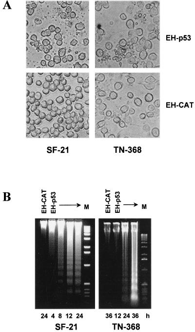
FIG. 5.
Expression of human p53 in insect cells induces typical apoptosis. (A) Light microscopy of SF-21 and TN-368 cells transfected with plasmids expressing EH-p53 or, as a negative control, EH-CAT. Photographs were taken at 12 h (SF-21) or 24 h (TN-368) after heat shock, using an Olympus IX50 microscope. (B) Insect cells were transfected with plasmids expressing the proteins indicated the lanes. Total DNA was harvested at various times after heat shock (indicated at the bottom) and analyzed by agarose gel electrophoresis. M, DNA molecular weight markers (1-kb DNA ladder).

