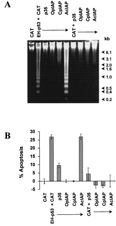
FIG. 6.
Inhibition of p53-induced cell death in insect cells by antiapoptotic genes. SF-21 cells were cotransfected with plasmids expressing the proteins indicated at the top. Cellular DNA was harvested 24 h after heat shock and examined by electrophoresis in a 1.2% agarose gel (A). Positions of DNA molecular weight markers (1-kb DNA ladder) are indicated at the right. The percentage of cells undergoing apoptosis was determined by trypan blue exclusion 24 h after heat shock (B). A plasmid expressing CAT was used as a negative control and to balance plasmid DNA concentrations. The percentage of apoptotic cells was calculated relative to the CAT control, set at 0%, which was similar to the level for mock-transfected controls.

