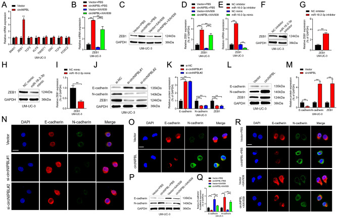
Fig. 5.
circNIPBL facilitates the expression level of ZEB1. (A) qRT-PCR analysis showed that the expressions of downstream targets of Wnt signaling pathway in circNIPBL overexpressing BCa cells. (B) qRT-PCR analysis of ZEB1 expression in indicated UM-UC-3 cells. (C, D) Representative images (C) and quantification (D) of Western blotting assay of ZEB1 in indicated UM-UC-3 cells. (E) qRT-PCR analysis of ZEB1 expression in indicated UM-UC-3 cells. (F-I) Representative images and quantification of Western blotting assay of ZEB1 in UM-UC-3 cells transfected with miR-16-2-3p inhibitors (F, G) and mimics (H, I). (J-M) Representative images and quantification of Western blotting assay of N-cadherin, E-cadherin, ZEB1 after circNIPBL knockdown (J, K) or circNIPBL overexpression (L, M) in UM-UC-3 cells. (N, O) The expression of N-cadherin and E-cadherin was detected by IF assay in circNIPBL knockdown (N) or circNIPBL overexpression (O) UM-UC-3 cells. Scale bar = 5 μm. (P, Q) Representative images (P) and quantification (Q) of Western blotting assay of N-cadherin and E-cadherin in indicated UM-UC-3 cells. (R) The expression of N-cadherin and E-cadherin was detected by IF assay in indicated UM-UC-3 cells. Scale bar = 5 μm. The statistical difference was assessed with one-way ANOVA followed by Dunnett tests in B, D, E, K and Q; and the two-tailed Student t test in A, G, I and M. Error bars show the SD from three independent experiments. *p < 0.05 and **p < 0.01

