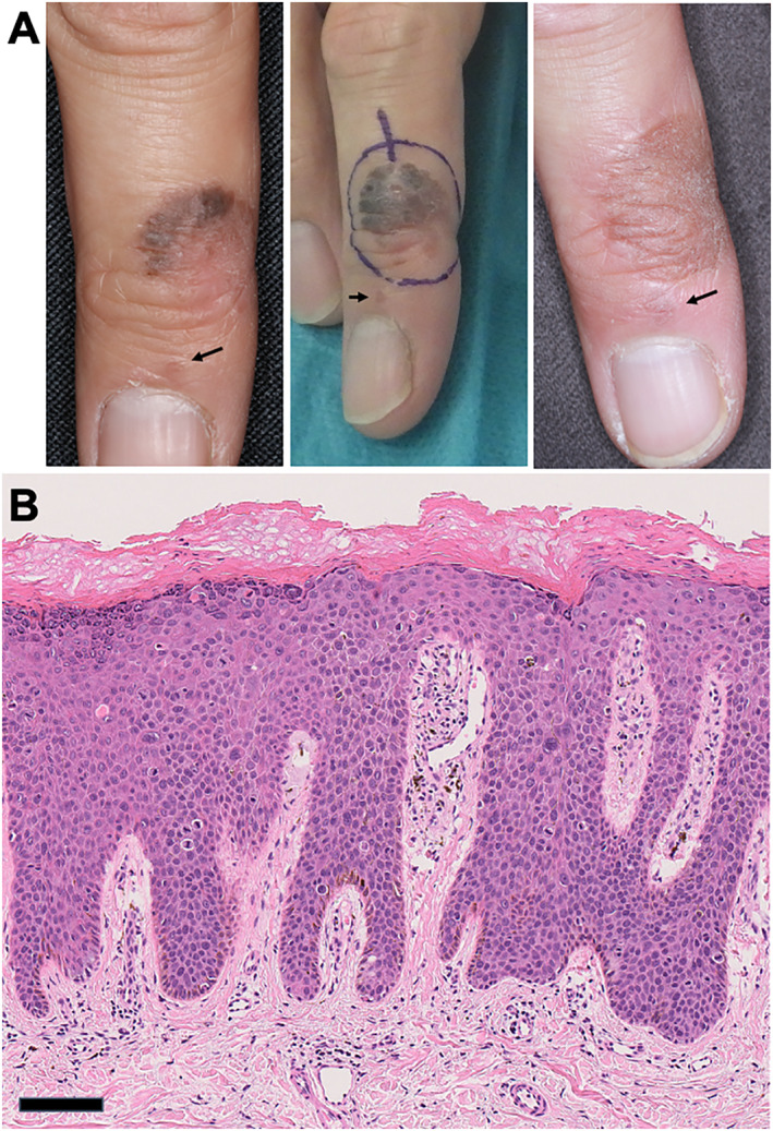
FIGURE 1.

Bowen's disease on the dorsal side of the right middle finger. (a) Clinical manifestation on the initial examination (left), excisional design (middle), and the manifestation 1 year after the first excision (right). Arrows represent the periungual lesions of Bowen's disease. (b) The histology shows that atypical basaloid cells replaced the full thickness of the epidermis with some clumping cells and individual cell keratinisation (haematoxylin and eosin staining, scale bar: 100 μm).
