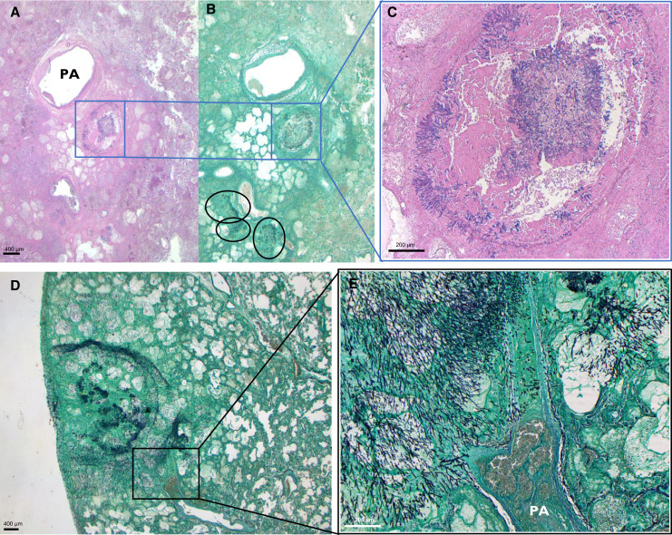
Figure 2.
Viral-associated pulmonary aspergillosis: unimpeded fungal growth pattern. (A–C) Histological images of a patient with influenza-associated pulmonary aspergillosis (IAPA). Histological image derived from left lower lobe, at a magnification of ×8.75; hematoxylin and eosin stain (A) and Grocott-Gomori’s methenamine silver stain (Grocott) (B). Multifocal unimpeded fungal growth within an area of coagulative necrosis is visualized, indicated by black circles and blue box. (C) Magnification of ×50 of the blue-boxed area, showing intrabronchial hyphal growth. (D and E) Lung slide Grocott staining of another patient with IAPA at ×10 (D) and ×50 (E), showing isometric centrifugal growth, invading into a bifurcating artery. PA = pulmonary artery.

