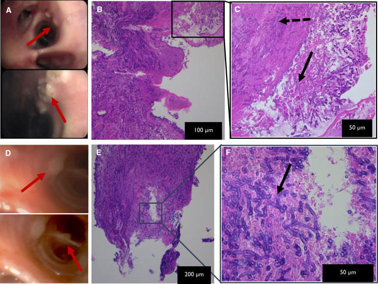
Figure 4.
Viral-associated invasive Aspergillus tracheobronchitis. Influenza-associated invasive Aspergillus tracheobronchitis: (A) Macroscopic image from bronchoscopy, showing extensive white nodular (red arrows) tracheobronchitis with central ulceration of noduli. (B and C) Microscopic hematoxylin and eosin–stained images at different magnifications, as indicated on the pictures of endobronchial biopsy, showing ulcerated epithelium with neutrophilic debris and acute-angle branching hyphae (black solid arrow) and hyphal invasion into tissue (black dashed arrow). Similar combination of macroscopic (D) and microscopic (E and F) evaluations from a case of coronavirus disease (COVID-19)–associated invasive Aspergillus tracheobronchitis. More extensive macroscopic inflammation visualized in influenza setting, yet comparable microscopic image in influenza and COVID-19 viral background.

