FIGURE 1.

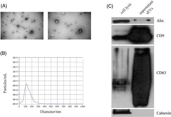
Verification of the identity of sEVs isolated from amniotic fluid. (A) Transmission electron microscopy (TEM) images of sEVs combined with SEC and UC. (B) NTA detection of sEVs enriched from AF, approximately 75‐200 nm in diameter. (C) EV markers CD9, CD63 and Alix detection in the sEVs isolated from amniotic fluid, and Calnexin, a negative marker of EV, was absent in our isolated sEVs.
