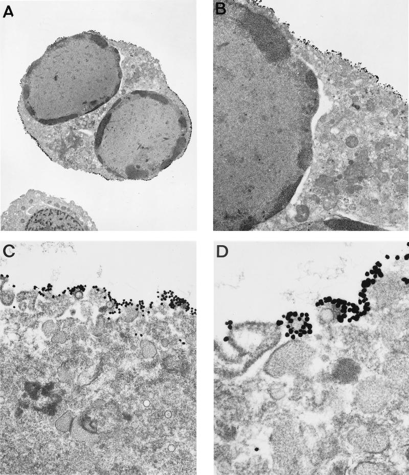
FIG. 4.
Immunoelectron microscopy of TPA-stimulated BCBL-1 cells with anti-K8.1 antibody. TPA-stimulated BCBL-1 cells were fixed, embedded in low-temperature resin, sectioned, and labeled with a combination of antigen-specific anti-K8.1 antibody and 5-nm-diameter colloidal gold. The sections were analyzed with a JEOL 1010 electron microscope. Magnifications: (A) ×3,600; (B) ×12,000; (C) ×36,000; (D) ×72,000.

