Figure 4.
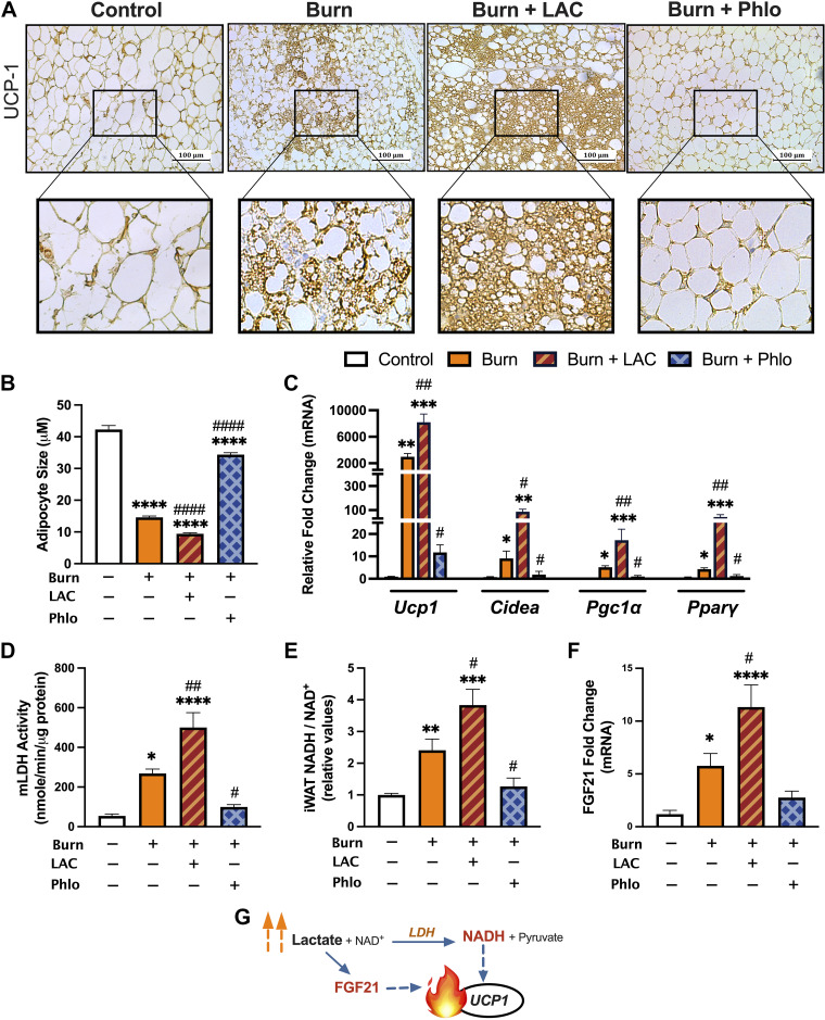
Increased MCT-mediated lactate shuttling augments postburn beige adipocyte formation. A: immunohistochemical staining for UCP1 in iWAT from control, vehicle-treated, l-lactate-treated, and Phloretin-treated burn mice at 7 days after injury. B: postburn changes in adipocyte diameter control (n = 116), vehicle-treated (n = 241), l-lactate-treated (n = 225), and Phloretin-treated (n = 160) burn mice at 7 days after injury. C: quantitative RT-PCR analysis of browning genes in inguinal WAT at 7 days postinjury. Changes in mitochondrial LDH activity (D), relative [NADH/NAD+] (E), and FGF21 gene expression (F) at 7 days postinjury. G: schematic depicting redox-dependent and -independent mechanisms of lactate-induced browning. Values are presented as means ± standard error. One-way ANOVA, Sham vs. burn *P < 0.05; **P < 0.01; ***P < 0.001; ****P < 0.0001, vehicle vs. treated burn #P < 0.05, ##P < 0.01, ####P < 0.0001, n = 7/group. WAT, white adipose tissue.

