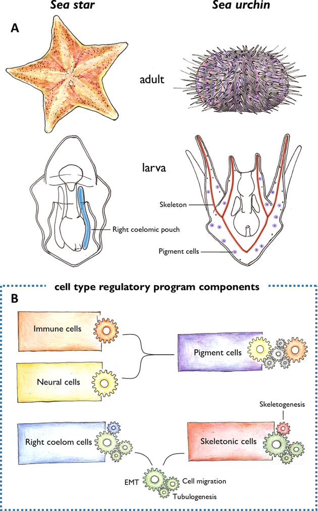
Figure 1. Morphological novelties in the sea urchin larva.
(A) Schematic representation of the adult (top) and larval (bottom) forms of sea stars and sea urchins. Two larval cell types had been thought to be unique to the sea urchin: the skeletogenic mesenchyme cells that make the skeleton (red), and the pigment cells embedded in the larval epithelium (purple). Meyer et al. found that the skeletogenic mesenchyme cells express similar genes to the right coelomic pouch of the sea star larva (blue), and the pigment cells are similar to immune and neural cells of the sea star larva (not shown). (B) These cells were found to share regulatory mechanisms (represented by different coloured clockwork wheels). Pigment cells activate similar regulatory components as immune (orange wheel) and neural (yellow wheel) cells in sea stars. Unknown components (grey wheels) may control the interplay between the immune and neural functions of these cells. The right coelomic pouch of the sea star turns on regulatory components required to produce mesenchymal cells (green wheels) – epithelial to mesenchymal transition (EMT), cell migration, and tubulogenesis – which are also activated in skeletogenic mesenchyme cells in sea urchins. This suggests that skeletogenic mesenchyme cells and cells of the right coelomic pouch evolved from the same ancestor. Later in evolution, the skeletogenic mesenchyme cells acquired a skeletogenic program (red wheel), while the right coelomic pouch cells might have acquired other currently unknown programs (blue wheel).
Image credit: Antonia Grausgruber.

