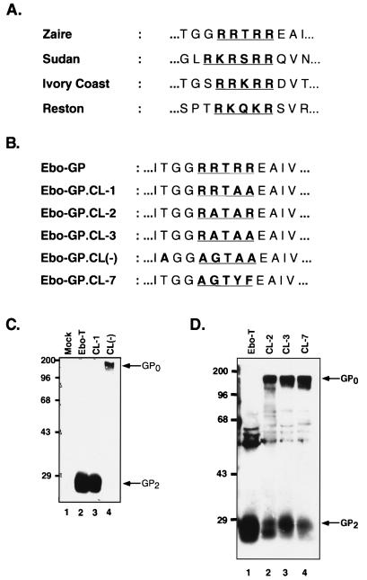
FIG. 3.
Identification of the Ebo-GP endoproteolytic processing site. (A) Comparison of the conserved dibasic motifs in the envelope glycoproteins of all known strains of Ebola virus. (B) Comparison of the Ebo-GP endoproteolytic processing site with those of five mutant glycoproteins. (C) Western blot analysis of Ebo-T, Ebo-T.CL-1, and Ebo-T.CL(−). 293T cells were transfected with either pCB6-Ebo-T (Ebo-T), pCB6-Ebo-T.CL-1 (CL-1), or pCB6-Ebo-T.CL(−) [CL(−)] or without a glycoprotein construct (Mock). Viral lysates prepared 48 h posttransfection were analyzed by SDS-PAGE and Western blotting with the anti-RSV tail serum. Arrows indicate the GP2 and GP0 proteins. Positions of molecular mass markers (in kilodaltons) are shown to the left of each gel. (D) Western blot analysis of Ebo-T, Ebo-T.CL-2, Ebo-T.CL-3, and Ebo-T.CL-7. Viral preparation and analysis were performed as for panel C. Arrows indicate the GP2 and GP0 proteins; positions of molecular mass markers (in kilodaltons) are shown to the left of each gel.

