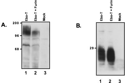
FIG. 5.
Expression of Ebo-T in LoVo cells. LoVo cells were transiently transfected with either no DNA (Mock), pCB6-Ebo-T (Ebo-T), or pCB6-Ebo-T and pGEM7hFurin (Ebo-T + Furin). Two hours posttransfection, the cells were infected with the vaccinia virus vTF-7. Eighteen hours postinfection, the cells were lysed and cellular protein expression was examined by SDS-PAGE and Western blotting with either the anti-Ebo-GP serum (A) or the anti-RSV tail serum (B). Positions of molecular mass markers (in kilodaltons) are shown to the left of each gel.

