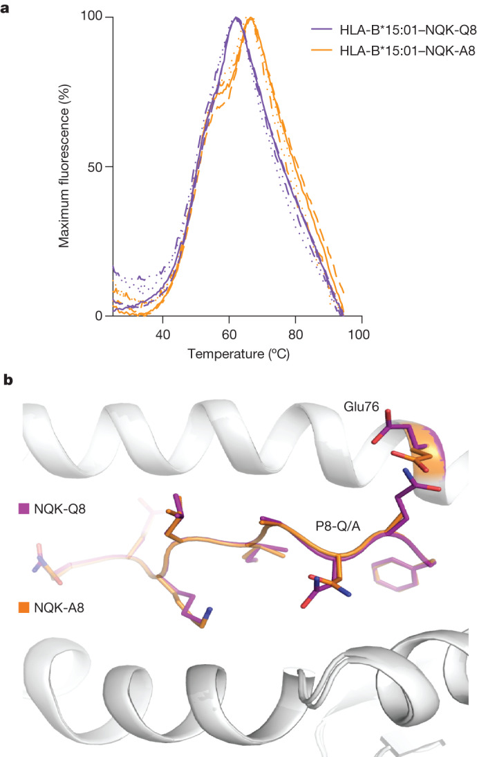
Fig. 4. NQK peptides are stable and adopt the same conformation bound to the HLA-B*15:01 molecule.

a, DSF plots showing the normalized fluorescence intensity versus temperature for HLA-B*15:01 in a complex with the NQK-Q8 (purple) or NQK-A8 (orange) peptide measured at concentrations of 5 μM and 10 μM. n = 2 biologically independent experiments performed in duplicate, represented by the different lines. b, Superimposition of the crystal structures of HLA-B*15:01 (white cartoon) in a complex with either the NQK-Q8 (purple stick) or the NQK-A8 (orange stick) peptide.
