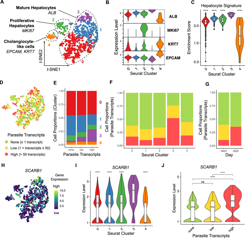
Fig. 2. Expression of hepatocyte maturation markers in Pf-infected HepOrgs at 4 or 5 days p.i.
A t-SNE map showing the Seurat clusters and major cell types in the human read dataset (n = 1277 cells; cluster 0, n = 571 cells; cluster 1, n = 426 cells; cluster 2, n = 117 cells; cluster 3, n = 100 cells; cluster 4, n = 63 cells). B Violin plot showing the lineage marker gene expression per Seurat cluster. C Violin plot showing the enrichment scores of the hepatocyte gene signature per Seurat cluster. D t-SNE map highlighting the infection rates of the cells (none, n = 786 cells; low, n = 269 cells; high, n = 222 cells). E Column chart showing the cell proportions per Seurat cluster and infection rate. Column chart showing the cell proportions per infection rate and F Seurat cluster or G collection day (4 or 5 days p.i.). H t-SNE map showing SCARB1 expression. Violin plots showing SCARB1 expression per I Seurat cluster and J infection rate. Two-sided Mann–Whitney U (Wilcoxon rank-sum) test: ns p ≥ 0.05, ****p < 0.0001. Statistics in C and I were calculated in comparison to cluster 3. C p < 2–16 (all comparisons); I p < 2–16 (vs cluster 0), p = 3.1–13 (vs cluster 1), p = 8.6–8 (vs cluster 2), p = 2.7–12 (vs cluster 4); J p = 0.53 (none vs low), p < 2.22–16 (none vs high), p = 1.9–14 (low vs high). Box plots in C, I and J indicate the median (Q2), 25th percentile (Q1) and 75th percentile (Q3) with the whiskers showing the minimum (Q1 – 1.5 × interquartile range) and maximum (Q3+ 1.5 × interquartile range). See also Fig. S2–S6, S11 and Supplementary Data 1. Source data are provided as a Source data file and Supplementary Data 5.

