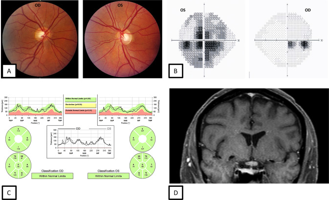
Fig. 1. 57-year-old male with AQP4-IgG positive neuromyelitis optica spectrum disorder (NMOSD) optic neuritis.
During the acute optic neuritis attack, fundus photographs demonstrate no optic disc oedema (A). Automated visual field testing shows a junctional scotoma (B). OCT shows a normal pRNFL thickness (C). Coronal, fat saturated T1-weighted post contrast MRI of the orbits shows enhancement of the optic chiasm and left optic nerve at the junction of the chiasm correlating with the visual field defects (D).

