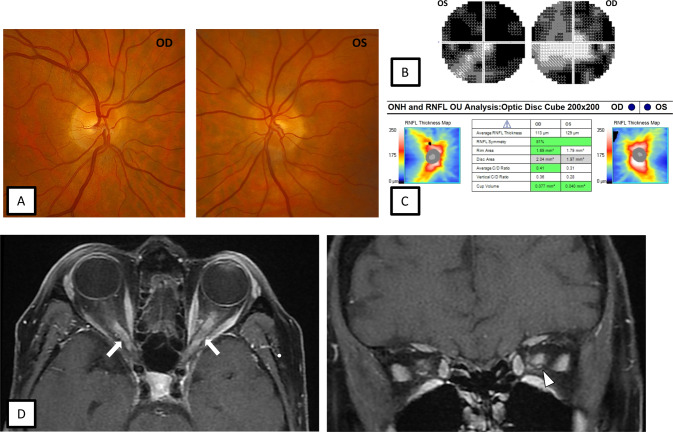
Fig. 2. 67-year-old female with myelin oligodendrocyte glycoprotein antibody-associated disease (MOGAD) optic neuritis.
During the acute optic neuritis attack, fundus photographs show trace bilateral optic disc oedema (A). Automated visual field testing demonstrates bilateral generalised constriction (B). OCT shows mild bilateral pRNFL thickening (C). Axial and coronal, fat saturated T1-weighted post contrast MRI of the orbits shows bilateral longitudinally optic nerve enhancement (white arrows) with mild perineural enhancement on the left (white arrowhead) (D).

