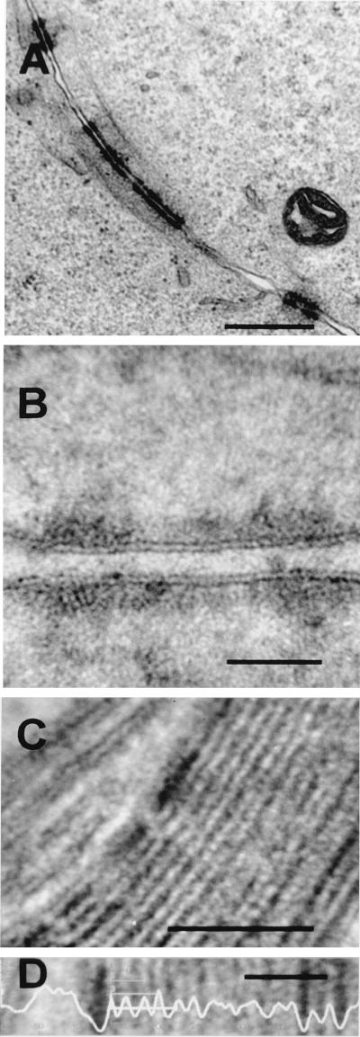
FIG. 5.
Tightly apposed cellular membranes at cell junctions (A and B) and in myelin sheaths around axons (C) remain discernible as individual trilaminar membranes. (A and B) HeLa cells were infected with VV strain WR, and at 24 h p.i. they were processed for electron microscopy as described in Materials and Methods. Images show a junction between cells. (C and D) Sample of guinea pig brain tissue processed for conventional electron microscopy, showing the myelin sheath surrounding the axon of a neuron. (D) Using image analysis (horizontal mean intensity profile) on a portion of the section shown in panel C, the periodicity between lipid bilayers remains 5.07 ± 0.81 to 5.11 ± 0.64 nm. See Table 2 for measurements of peak-to-peak distances. Bars, 500 nm (A), 50 nm (B and C), and 25 nm (D).

