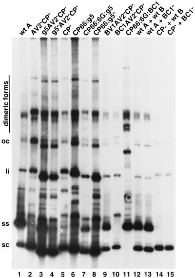
FIG. 2.
Replication of ToLCV constructs in infected BY2 protoplasts. Southern blot analysis was performed as described in Materials and Methods. The viral constructs used for infecting protoplasts are shown above the lanes. Protoplasts were inoculated with A-component DNA alone (lanes 1 to 11) or coinoculated with A- and B-component DNAs (lanes 12 to 15). Each lane contained 4 μg of DNA prepared from protoplasts (single transfection). Viral DNA was detected with a radioactively labeled probe from A-component DNA. The positions of supercoiled (sc), single-stranded (ss), linear (li), and open circular (op) viral DNA forms are indicated. Note that the positions of supercoiled and other viral DNA forms in lane 11 are shifted upward due to the larger size of the CP66:6G:BC1 construct. wt, wild type.

