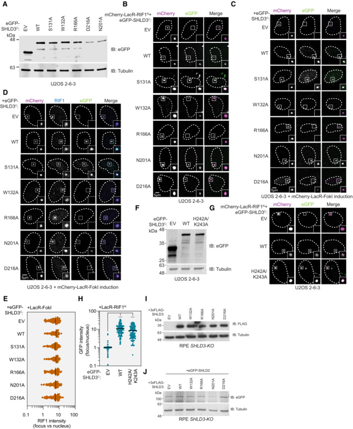
Figure EV3. Data supporting the polar interactions that are essential for SHLD3‐RIF1 binding.

- Immunoblot of whole cell extracts of U2OS 2‐6‐3 cells transfected with plasmids encoding the indicated eGFP‐tagged SHLD3C (residues 126–250) variants. Lysates were probed for eGFP and tubulin (loading control). IB—immunoblot. EV—empty vector. WT—wild‐type.
- Representative micrographs of the LacR/LacO assay using mCherry‐LacR‐RIF1N as bait to evaluate chromatin recruitment of eGFP‐tagged SHLD3C alanine substitution variants shown in Fig 3C. SHLD3C: residues 126–250. RIF1N: residues 1–967.
- Representative micrographs of the LacR‐FokI assay to evaluate DNA‐damage recruitment of eGFP‐tagged SHLD3C alanine substitution variants after induction of LacR‐FokI expression shown in Fig 3D. SHLD3C: residues 126–250.
- Representative micrographs of the LacR‐FokI assay to evaluate DNA‐damage recruitment of endogenous RIF1 after induction of LacR‐FokI expression in the presence of exogenously expressed eGFP‐tagged SHLD3C alanine substitution variants. SHLD3C: residues 126–250.
- Quantification of D. RIF1 immunofluorescence intensities are presented as a ratio between the average fluorescence intensity within the mCherry‐labeled LacR focus and the average nuclear intensity. Bars represent mean values (n = 99, 92, 88, 97, 90, 92, 100 for EV, WT, S131A, W132A, R166A, N201A, D216A from two biologically independent experiments).
- Immunoblot of whole cell extracts of U2OS 2‐6‐3 cells transfected with plasmids encoding the indicated eGFP‐tagged SHLD3C (residues 126–250) variants. Lysates were probed for eGFP and tubulin (loading control).
- Representative micrographs of the LacR/LacO assay using mCherry‐LacR‐RIF1N as bait to evaluate chromatin recruitment of eGFP‐tagged SHLD3 H242A/K243A.
- Quantification of G. GFP intensities are presented as a ratio between the average fluorescence intensity within the mCherry‐labeled LacR‐RIF1N focus and the average nuclear intensity. Bars represent mean values (n = 127, 129, 136 for EV, WT, H242A/K243A from three biologically independent experiments). Analysis was performed using the Kruskal–Wallis test followed by Dunn's multiple comparisons against empty vector control. ****P < 0.0001.
- Immunoblot of whole cell extracts of RPE SHLD3‐KO cells stably transduced with lentivirus encoding the indicated 3xFLAG‐tagged SHLD3 alanine substitution variants. Lysates were probed for FLAG and tubulin (loading control). IB—immunoblot.
- Immunoblot of whole cell extracts of complemented RPE SHLD3‐KO cells shown in (I) that were transfected with plasmid encoding eGFP‐SHLD2. Lysates were probed for eGFP and tubulin (loading control).
Source data are available online for this figure.
