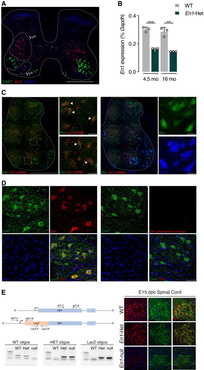
Figure 1. En1 expression in the adult spinal cord.

- Triple RNAscope in situ hybridization showing Engrailed‐1 (En1), Choline acetyltransferase (ChAT), and Calbindin‐1 (Calb1) expression in the lumbar spinal cord. En1 is expressed in V1 interneurons, dorsal (V1P) and ventral (V1R) to the main ChAT‐expressing motoneuron pool. Ventral interneurons correspond to Renshaw cells as shown by Calb1 expression. Scale bar: 500 μm.
- RT–qPCR of RNA from the lumbar enlargement at 4.5 and 16 months of age shows stable En1 expression in WT at both ages and a twofold reduction of expression in heterozygous mice. Unpaired two‐sided t‐test. **P < 0.005; ***P < 0.0005. n = 3. Values are mean ± SD.
- Triple staining EN1 IHC (green), En1 RNAscope ISH (red), and ChAT RNAscope (blue) demonstrating the double staining of En1 mRNA and protein (EN1) in the V1 interneuron population (left panel insets, arrowheads point toward examples of double‐stained V1 interneurons), and the presence of EN1 protein in large cells not expressing En1 mRNA (left panel) but expressing ChAT (right panel insets). Scale bar: 500, 30 μm for high magnification insets.
- Left: EN1 (red) detected with the LSBio antibody is localized in ChAT‐expressing neurons (green) in the ventral horns of the spinal cord. Right: EN1 signal is lost upon preincubation of the antibody with 1.5 M excess of recombinant hEN1. Scale bar: 50 μm.
- Left: Relative positions of the different oligonucleotides selected to genotype the E15 embryos and examples of the genotyping based on the combination of PCRs with the different pairs of primers. Right: Double ChAT/EN1 immunostaining demonstrating the co‐localization of the two proteins in the WT and the En1‐Het ventral cord and the absence of EN1 staining in MNs from En1‐KO embryos. Note that the staining is reduced in En1‐Het embryos, compared with WT embryos.
Data information: These experiments were performed once.
Source data are available online for this figure.
