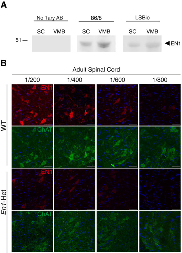
Figure EV1. Characterization of the anti‐EN1 LSBio antibody.

- Western blots of spinal cord (SC) and ventral midbrain (VMB) extracts demonstrating that the 86/8 and LSBio antibodies recognize in both structures the same protein migrating with recombinant EN1 velocity. No staining is observed in the absence of primary antibody (left panel). This experiment was performed twice.
- Double staining of 3‐month‐old WT and En1‐Het ventral MNs with the anti‐ChAT antibody and the anti‐EN1 LSBio antibody at various dilutions. EN1 staining decreases with increasing dilutions of the antibody. The loss of staining is more rapid in En1‐Het than in WT mice. Scale bar = 50 μm.
Data information: This experiment was performed once.
