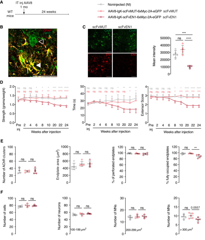
Figure 4. Effects of extracellular EN1 neutralization on strength, MN survival, and NMJ morphology.

- Experimental paradigm and structure of AAV8‐encoded constructs containing glial fibrillary acidic protein (GFAP) promoter for expression in astrocytes, Immunoglobulin K (IgK) signal peptide for secretion, anti‐ENGRAILED single‐chain antibody (scFvEN1), 6 myc tags (6xMyc), skipping P2A peptide, and enhanced Green Fluorescent Protein (eGFP). An inactive control antibody (scFvMUT) contains a cysteine to serine mutation that prevents disulfide bond formation between IgG chains, thus epitope recognition. The AAV8 was injected in 1‐month‐old WT mice, and the strength phenotypes were followed for 6 months before anatomical analysis.
- Analysis of 1‐month postinjection showing that scFv antibodies are expressed in astrocytes (white arrowhead) double‐stained for GFAP and Myc and exported (empty arrowhead). Scale bar: 50 μm.
- Left panel illustrates that expressing the scFvEN1, but not scFvMUT, abolishes EN1 staining by LSBio anti‐EN1 antibody in ventral horn ChAT+ cells and right panel quantifies this inhibition 1‐way ANOVA followed by Tukey corrected post hoc comparisons (n = 5 mice per group, ***P < 0.0005, ****P < 0.0001). Scale bar: 100 μm.
- The three graphs illustrate how the WT antibody but not its mutated version leads to progressive strength decrease. Two‐way ANOVA showed significant main effects for grip strength (F(2, 12) = 15.88, P < 0.0005), inverted grid (F(2, 107) = 19.86, P < 0.0001), and extensor score (F(2, 12) = 30.22, P < 0.0001) followed by Tukey corrected post hoc comparisons (*P < 0.05; **P < 0.005; ***P < 0.0005; ****P < 0.0001). n = 5 per treatment.
- Six months following infection (7‐month‐old mice), extracellular EN1 neutralization does not modify the number of AChR clusters, nor the endplate surface area, nor the percentage of perforated endplates. In contrast, the percentage of fully occupied endplates is diminished (right end panel). 1‐way ANOVA followed by Tukey corrected post hoc comparisons (**P < 0.005). n = 5 per treatment.
- Six months following infection, extracellular EN1 neutralization does not globally modify the total neuron number of cells at the lumbar level (left panel). A separate analysis of small (100–199 μm2), medium (200–299 μm2), and large (>300 μm2) neurons demonstrate a specific (P < 0.0557) loss of the latter category (αMNs). 1‐way ANOVA followed by Tukey corrected post hoc comparisons. N = 5 per treatment.
Data information: The extracellular neutralization study was performed twice. Values are mean ± SD.
Source data are available online for this figure.
