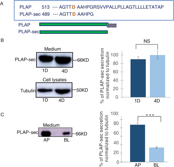
FIGURE 1:
PLAP-sec is apically secreted in polarized MDCK cells. (A) Schematic representation of secretory PLAP-sec. (B) MDCK cells stably expressing PLAP-sec (MDCK PLAP-sec) cultured for 1 d (1D) or 4 d (4D) were left to secrete for 4 h before cellular media were collected to monitor PLAP-sec secretion. In parallel, intracellular tubulin levels were monitored. On the right quantification of PLAP-sec secretion in 1D and 4D normalized to intracellular tubulin in four different experiments is shown. No statistical differences were revealed. (C) MDCK PLAP-sec cultured for 4 d on filters were left to secrete for 4 h before the cellular media were collected to further monitor apical (AP) or basolateral (BL) PLAP-sec secretion. On the right, quantification from three different experiments, ***p < 0.001.

