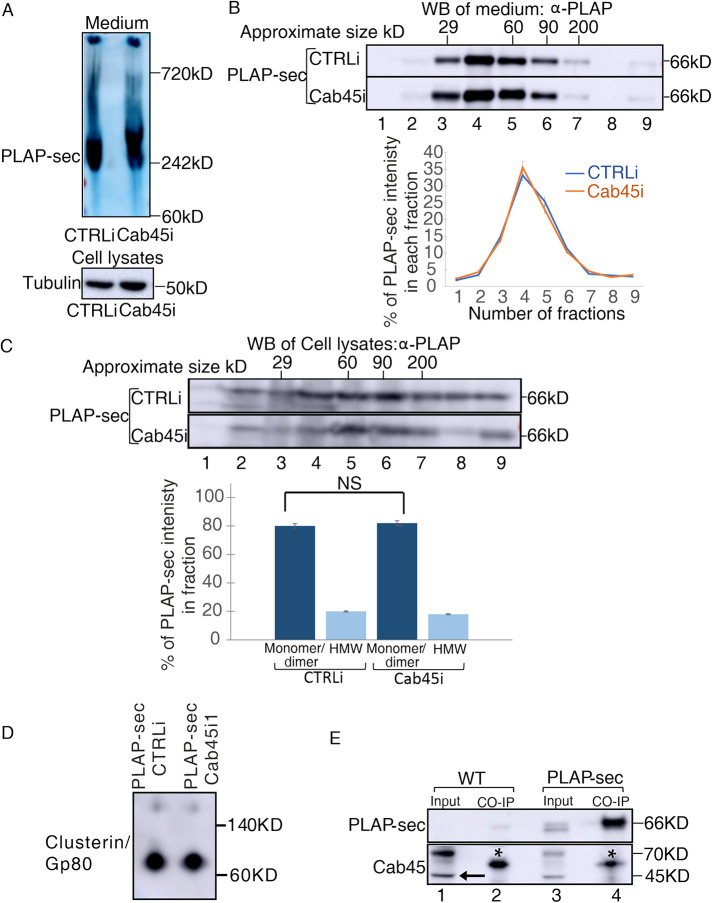
FIGURE 3:
The loss of Cab45 does not affect secreted and intracellular PLAP-sec clustering. (A, B) Cellular media of MDCK PLAP-sec CTRLi and Cab45i cells plated for 4 d were analyzed by (A) Native PAGE or (B) velocity gradient where fractions collected from top (fraction 1) to bottom (fraction 9) were loaded on SDS–PAGE gel to monitor clustering of secreted PLAP-sec. The representative molecular weight markers are indicated. Native-PAGE experiments were performed three times and velocity gradient five times with the quantification shown on the right. (C) Cell lysates of MDCK PLAP-sec CTRLi or Cab45i cells seeded for 4 d were collected and run on velocity gradient (method). Fractions were collected and then trichloroacetic acid (TCA) precipitated and analyzed by SDS–PAGE with PLAP antibody. The experiment was repeated four times, and the quantification is shown. (D) Cell lysates of MDCK PLAP-sec CTRLi or Cab45i seeded for 4 d were run on Native PAGE to monitor the Gp80/clusterin. (E) Cell lysates of MDCK PLAP-sec or MDCK WT cells seeded for 4 d were immunoprecipitated with PLAP antibody together with the protein A Sepharose beads and revealed with either anti-PLAP (top) or anti-Cab45 (bottom) antibodies. Aliquots of cell lysates (1: input; 2: pull down) were loaded. *, most probably heavy chain of immunoglobulin G. Note that no interaction could be monitored between PLAP-sec and Cab45.

