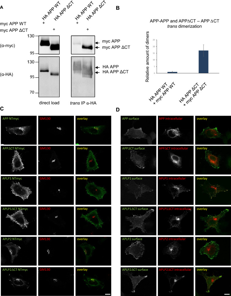
Fig. 4.
Trans-dimerization of the APP gene family members lacking the C-terminus. A Trans-co-immunoprecipitation of APP WT or APP∆CT, trans-homodimers. N2a cells were transiently transfected in separate dishes with myc APP, HA APP, myc APP∆CT or HA APP∆CT constructs. 4 h post-transfection, the following cells of separate dishes were combined: myc APP and HA APP, myc APP∆CT and HA APP∆CT. Equal amounts of cell lysates were loaded directly on an SDS gel and analyzed via Western blot with primary α-HA or α-c-myc antibodies (input controls). Further, equal amounts of cell lysates were used for immunoprecipitation with α-HA antibody coated agarose beads. The samples were separated on an SDS gel and subjected for Western blot detection with the primary antibody α-c-myc to detect the co-immunoprecipitated proteins. The same membrane was incubated afterwards with an α-HA antibody to detect total amounts of immunoprecipitated HA APP HA or HA APP∆CT. B Quantification of data shown in A. Bars represent mean values ± SEM; n = 3, unpaired Student’s t test *p < 0.05, **p < 0.01, ***p < 0.001. C HeLa cells were transiently transfected with N-terminally c-myc tagged APP, APP∆CT, APLP1, APLP1∆CT, APLP2 and APLP2∆CT. Cells were stained with α-c-myc antibody to visualize the overexpressed proteins and GM130 antibody to show the cis-Golgi apparatus in permeabilized cells. APP gene family members lacking the C-terminus are located to a higher amount at the cell surface. Scale bar, 10 µm. D Cell surface staining of HeLa cells transiently transfected with N-terminally c-myc tagged APP, APP∆CT, APLP1, APLP1∆CT, APLP2 or APLP2∆CT. Cells were incubated with an α-c-myc antibody on ice to stain only proteins which were localized at the surface. After fixation, the cells were permeabilized and stained again with an α-c-myc antibody to also visualize the intracellular proteins

