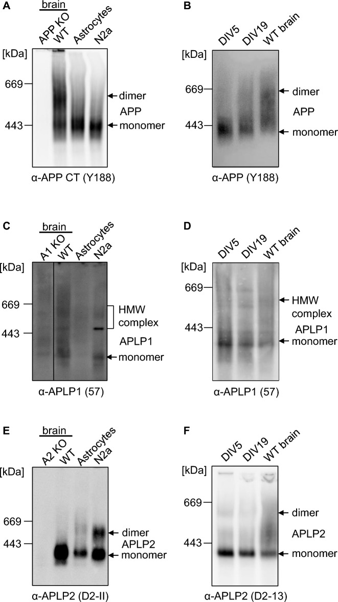
Fig. 8.
Higher amount of APP/APLP1/APLP2 dimerization is not visible in astrocytes, N2a cells or neurons. A, C, E One year old WT mouse cortices and APP KO mouse cortices were homogenized and compared with the lysate of astrocytes and N2a cells. All samples were prepared under semi-denaturing conditions for analysis on Blue Native gels. Western blot detection of APP followed with the primary α-APP C-terminal antibody Y188 (A), of APLP1 with the primary α-APLP1 antibody 57 (C) and APLP2 with the primary α-APLP2 antibody D2-II (E). B, D, F Mouse primary cortical neurons were cultivated for 5 or 19 days in vitro (DIV), lysed and compared to the lysate of 1 year old WT mouse cortices. All samples were prepared under semi-denaturing conditions for analysis on Blue Native gels. Western blot detection of APP followed with the primary α-APP C-terminal antibody Y188 (B), of APLP1 with the primary α-APLP1 antibody 57 (D) and APLP2 with the primary α-APLP2 antibody D2-II (F)

