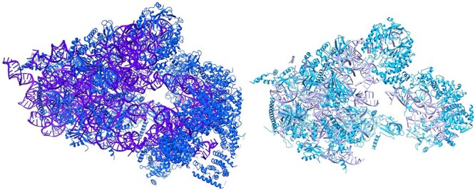
Figure 8.

A visualization of DeepTracer’s ribosome sample and EMD-32801 solved model. The left model is the solved structure, with blue for amino acids and purple for nucleotides. The right model is the DeepTracer version, with a light blue for amino acid and mauve for nucleotides.
