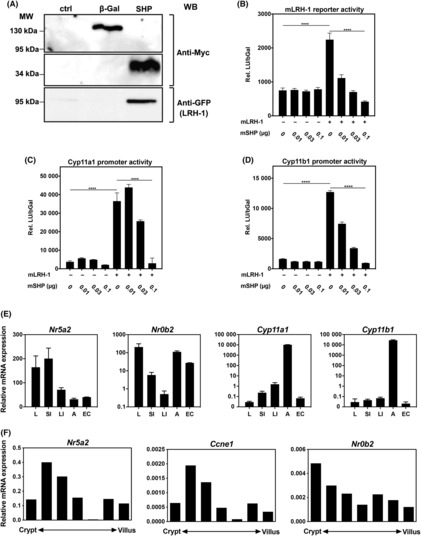
Fig. 2.

SHP suppresses LRH‐1 transcriptional activity. (A) Murine mICcl2 cells were co‐transfected with Myc/His‐tagged β‐Galactosidase (β‐Gal) as control or SHP, and GFP‐tagged LRH‐1. Nuclear lysates were precipitated with Ni2+‐Sepharose. Proteins were detected using anti‐Myc and anti‐GFP antibodies. (B–D) mICcl2 cells were transfected with an LRH‐1 luciferase reporter (B), Cyp11a1 promoter reporter (C) or Cyp11b1 promoter reporter (D), mLRH‐1 and increasing concentrations of mSHP expression plasmids. Relative luciferase units, normalized to β‐galactosidase activity (rel. LU/bGal) were assessed. Mean values of triplicates ± SD are shown. One‐way ANOVA with Turkey's multiple comparison ****P < 0.0001. (E) Detection of LRH‐1 (Nr5a2), SHP (Nr0b2), Cyp11a1 and Cyp11b1 mRNA expression by RT‐qPCR in liver, small intestine, large intestine, adrenal glands and isolated intestinal epithelial cells from wild‐type C57Bl/6 mice. Mean values of n = 3 ± SD are shown. (F) Expression of LRH‐1 (Nr5a2), cyclin E1 (Ccne1) and SHP (Nr0b2) along the crypt–villus axis. Epithelial cells from villus to crypt were differentially isolated and mRNA expression was analysed by RT‐qPCR. A typical experiment of n = 3 is shown.
