Fig. 3.

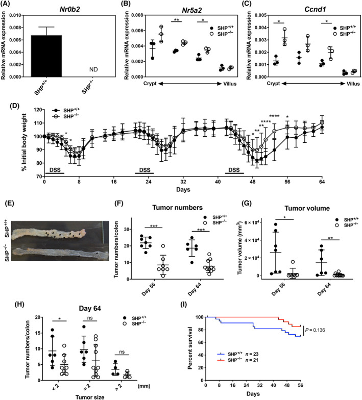
Effects of Shp deletion on LRH‐1‐regulated intestinal tumour development. (A) Detection of Shp (Nr0b2) in colonic tissue from control mice (Shp+/+) and Shp‐deficient mice (Shp−/−) (n = 3 mice per group, mean ± SD). ND, not detected. (B, C) Expression of Lhr‐1 (Nr5a2) and cyclin D1 (Ccnd1) along the crypt–villus axis. Epithelial cells from villus to crypt were differentially isolated from Shp+/+ and Shp−/− mice and mRNA expression was analysed by RT‐qPCR. Mean values ± SD of three mice per group are shown. (D) Weight loss curve of control (Shp+/+) and Shp−/− after AOM/DSS treatment. Mean values ± SD of pooled three independent experiments are shown (n = 7–14 mice per group). (E) Colonic tumour development (arrows) in control (Shp+/+) and Shp−/− at day 64. (F, G) Tumour numbers (F) and tumour volume (G) of Shp+/+ and Shp−/− mice at days 56 and 64 (mean ± SD of n = 7 mice per group). (H) Numbers of colonic tumours stratified according to size. Unpaired Student's t‐test, *P < 0.05, **P < 0.01, ***P < 0.001, ****P < 0.0001. (I) Overall survival of Shp+/+ and Shp−/− mice during AOM/DSS‐induced colitis.
