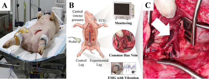
Fig 1. Experimental environment.
(A) Each pig was placed in a supine position on a surgical table. (B) The vital signs were continuously monitored and the electromyography (EMG) electrode, accelerometer, and vibration motor were attached to the tibialis anterior muscle of the right (control) and left (experimental) hind limb using straps. (C) A common iliac vein was identified in the retropelvic space (white arrow) for the left hind limb of the pig.

