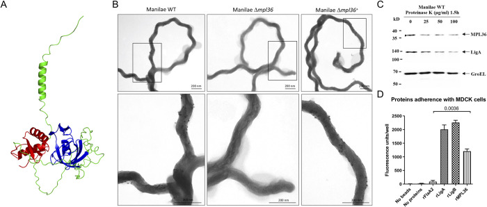
Fig 1. Structure and surface localization of MPL36 in L. interrogans serovar Manilae L495 and rMPL36 interaction with host epithelial cells.
(A) Predicted model of L. interrogans MPL36 with the double-psi beta-barrel domain (DPBB) (blue), a conserved region of RlpA proteins (green), and the SPOR domain (red) in the C-terminal. (B) Immunogold labeling of WT, Δmpl36, and Δmpl36+ Leptospira strains were performed using polyclonal rabbit antiserum against MPL36 and goat anti-rabbit IgG labelled with 10 nm colloidal gold particles. Cells were visualized using 2% UA negative staining. (C) Whole intact spirochetes were incubated with different concentrations of Proteinase K (25–100 μg/mL), and western-blot analysis was conducted using polyclonal rabbit antisera against MPL36, LigA (positive control), and GroEL (negative control). (D) Recombinant proteins coated with fluorescent latex beads were incubated with immobilized MDCK cells and this interaction was assessed by fluorescent emission. LigA and LigB were used as positive control, while FlaA2 was used as negative control. The results are represented as mean ± standard deviation of two independent experiments.

