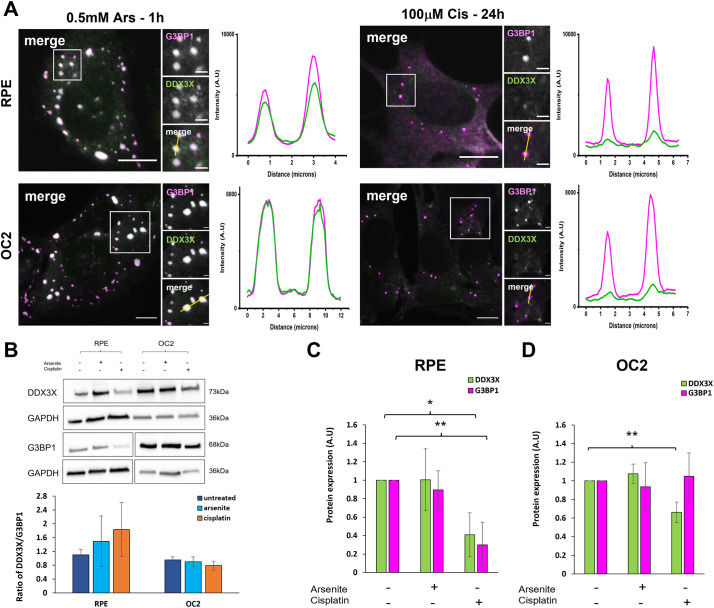
Fig. 5.
Cisplatin-induced SGs fail to sequester DDX3X effectively. (A) Representative widefield immunofluorescent images of RPE and OC2 cells treated with either 0.5 mM sodium arsenite for 1 h or 100 µM cisplatin for 24 h before fixation. Cells were immunostained for G3BP1 (magenta) and DDX3X (green). Fluorescence intensity line plots for G3BP1 and DDX3X were measured from the corresponding yellow line (in small merge panel). Scale bars: 10 µm (main images), 2 µm (smaller images). (B) Western blot of DDX3X and G3BP1 in untreated, arsenite and cisplatin treated cells and ratio of DDX3X to G3BP1 levels from densitometry. GAPDH was used as loading control for densitometry analysis of RPE (C) and OC2 (D) cells. Error bars represent s.d., n=4 experiments. *P<0.05, **P<0.01 (unpaired two-tailed Student’s t-test). A.U., arbitrary units.

