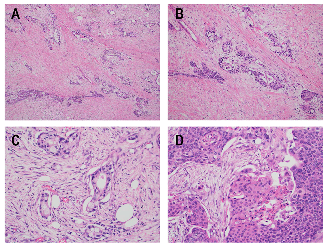
Figure 1. Histologic appearance of ASCP.

H&E specimens of ASCP. A) Areas resembling conventional ductal adenocarcinoma of the pancreas (top right) and squamous cell carcinoma (bottom left) are apparent in this sample (100x). Note the striking amount of desmoplastic stroma present between tumor nests. B) In this area of the tumor, the glandular and squamous components have merged together so that both morphologies can be seen within a single group of tumor cells (100x). C) Infiltrative glands making up the adenocarcinoma component of the tumor are shown at higher power (200x), with some cells containing possible mucin vacuoles. D) Solid sheets of tumor cells with densely eosinophilic cytoplasm and areas of keratinization, consistent with the squamous cell carcinoma component of the tumor, are shown at higher power (200x).
