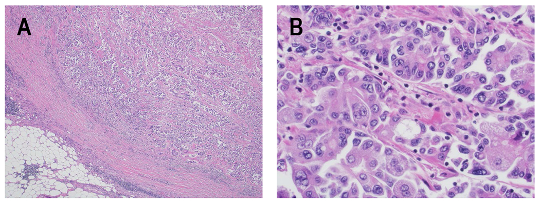
Figure 3. Histologic appearance of PACC.

H&E specimens of PACC. A) Low power image showing how PACC recapitulates the architecture of the exocrine pancreatic parenchyma with back-to-back acinar structures containing lumina. The tumor clusters are irregular in size and shape and are approaching the peri-pancreatic adipose tissue (40x). B) Higher power image showing the classic cytologic features of PACC with basally located nuclei containing single prominent nucleoli, and abundant apical amphophilic and granular cytoplasm (400x).
