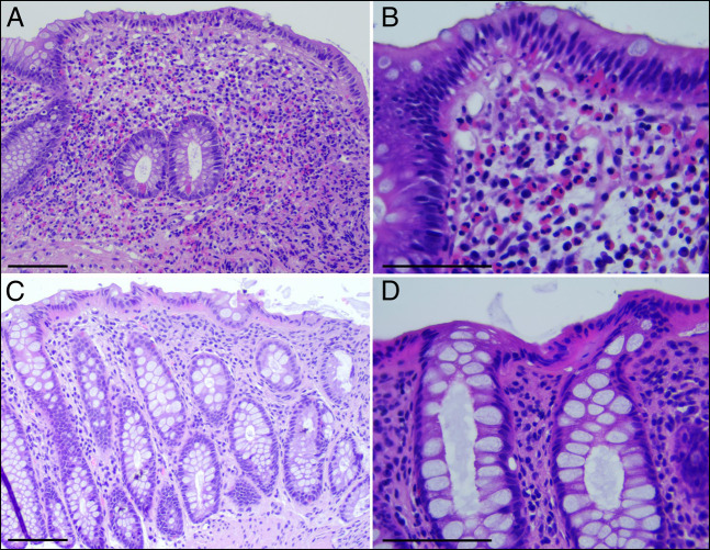
Figure 2.
(A and B) A hematoxylin and eosin-stained section of the large bowel mucosa obtained from the first colonoscopy. Biopsies are from the transverse colon showing numerous eosinophils in the lamina propria as well as in the surface and crypt epithelium. Scale bar 50 μm (A) and 25 μm (B). (C and D) Hematoxylin and eosin-stained sections of biopsies from the descending colon obtained during the second colonoscopy, after clopidogrel suspension, showing marked reduction of lamina propria eosinophils; these are rare and are never associated with crypts or surface epithelium. Scale bar 50 μm (A) and 25 μm (B).

