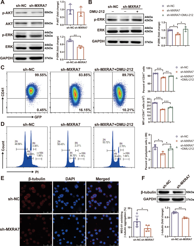
Figure 6.
Knock-down of MXRA7 inhibited the ERK/MAPK signaling pathway and decreased the expression of β-tubulin. MEG-01/sh-NC and MEG-01/sh-MXRA7 cells were incubated for 72 hours in the presence of 10 ng/mL rhTPO. (A) Proteins were extracted from cell lines for western blot analysis of p-AKT, AKT, p-ERK and ERK. (B) Proteins were extracted from cell lines treated with or without DMU-212 for western blot analysis of p-ERK and ERK. (C and D) Treated with or without DMU-212, the percentage and mean fluorescence (MFI) of CD41+ cells, and the percentage of polyploid cells (≥8N) were analyzed by flow cytometry. The expression of β-Tubulin was detected by immunofluorescence staining (E) and western blot (F). *P <.05, **P <.01, ***P <.001.

