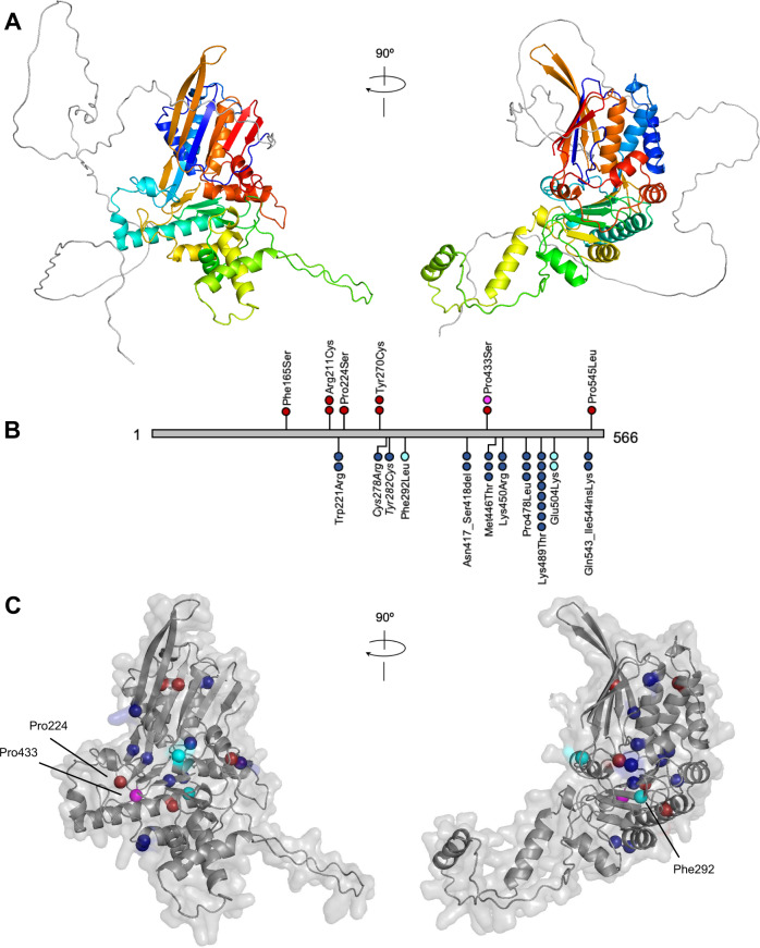
Fig. 2. Predicted structure of human DONSON and the locations of DONSON-MGORS and MISSLA missense variants.
A Predicted structure of human DONSON (AlphaFoldDB ID: DONS_HUMAN). DONSON is composed of a 150-residue long intrinsically disordered N-terminal region (grey) followed by interfolded protein (residues 151-566) coloured rainbow from the N-terminal end (blue) to the C-terminal end (red). B Linear DONSON (grey) showing the location of missense variants found in MGORS (above) and MISSLA (below). Each sphere represents one individual: red - MGORS, blue - MISSLA, magenta - MISSLA individual with clear MGORS features, cyan - MISSLA patients with phenotypic overlap to MGORS. Variants Cys278Arg/Tyr282Cys are italicized as either one, or both, are pathogenic in a single individual. C Position of MGORS and MISSLA-affected residues within the predicted DONSON structure. Residues are coloured by patient phenotype as per B. Indicated residues are discussed further in the main text. No variants are in the 150-residue-long intrinsically disordered N-terminal region and so for clarity it was removed.

