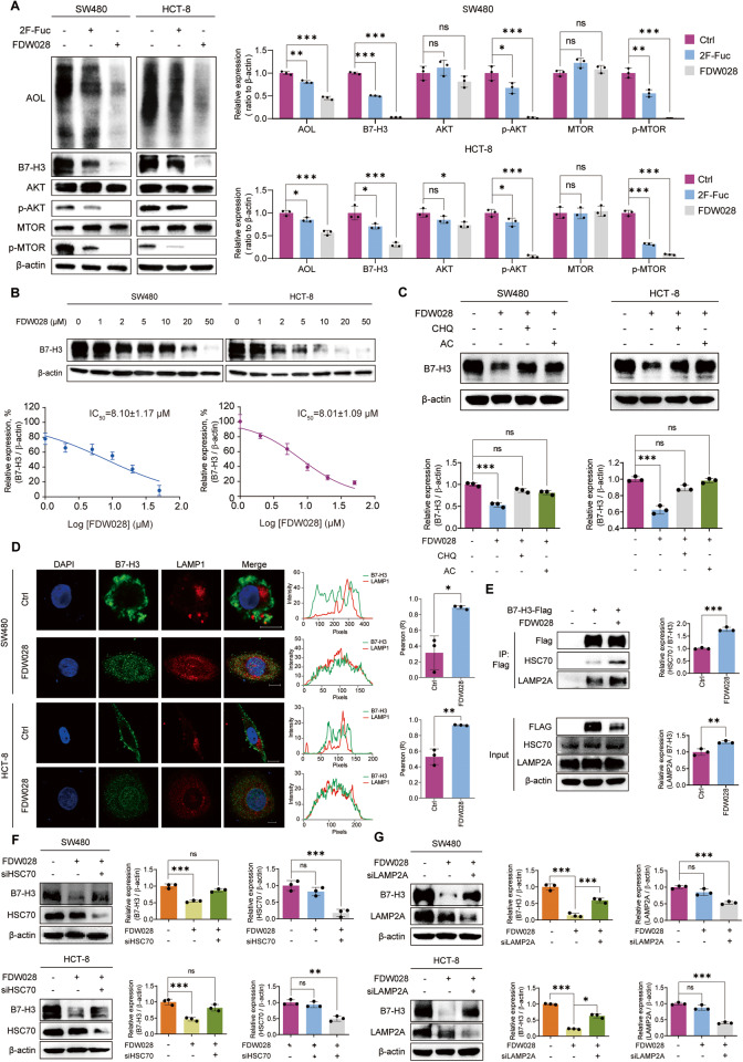
Fig. 6. FDW028 promoted defucosylation and CMA degradation of B7-H3.
A Immunoblots of AOL, B7-H3, and AKT/MTOR pathway molecules in SW480 and HCT-8 cells treated with FDW028 or 2F-Fuc at 50 μM for 72 h. B Immunoblots of B7-H3 in SW480 and HCT-8 cells treated with FDW028 at the indicated concentrations for 72 h. C Immunoblots of B7-H3 in SW480 and HCT-8 cells after the treatment of FDW028 (50 μM) with or without following CHQ (50 μM) or AC (100 μM). D Immunofluorescence shows the colocalization of B7-H3 and LAMP1 upon the treatment of FDW028. The intensity profiles of B7-H3 and LAMP1 are plotted in the middle panel. The statistical results of colocalization factor (Pearson’s R value) are shown on the right panel. E Co-IP assays showed the effects of FDW028 on the interaction between B7-H3 and HSC70 or LAMP2A. F Immunoblots of B7-H3 in SW480 and HCT-8 cells treated by FDW028 (50 μM) with or without HSC70 siRNA for 72 h. G Immunoblots of B7-H3 in SW480 and HCT-8 cells treated by FDW028 (50 μM) with or without LAMP2A siRNA for 72 h. All experiments were performed in technical triplicates and are displayed as mean ± s.d.; ns no significance; *P < 0.05, **P < 0.01, ***P < 0.001.

