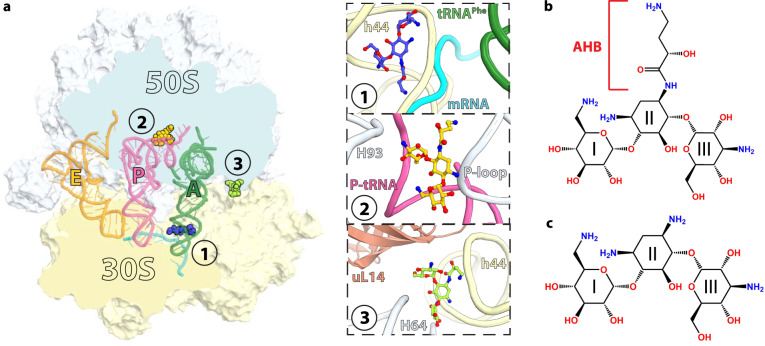
Fig. 1. Overview of the Thermus thermophilus 70S ribosome bound to amikacin.
a Overview of the 70S ribosome complexed with mRNA (cyan), tRNAPhe in the aminoacyl (A) site (green), tRNAifMet in the peptidyl (P) site (pink), tRNAPhe in the exit (E) site (orange), and AMK bound to three potentially relevant sites. (Inset 1) AMK bound near the decoding center in the small subunit, (inset 2) AMK bound in the large subunit P site, and (inset 3) AMK bound at inter-subunit bridge B5. Chemical structures of the antibiotics AMK with the amino-hydroxy butyrate (AHB) moiety at the N1 position of the central 2-deoxystreptamine (2-DOS) ring II (b), and KAN (c).

